
Ronald A. Bergman, Ph.D., Adel K. Afifi, M.D., Paul M. Heidger,
Jr., Ph.D.
Peer Review Status: Externally Peer Reviewed

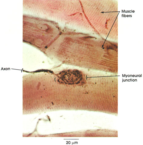
Rabbit, Ranvier's gold chloride method, 612 x.
The method used in this preparation is a classical technique for staining nerve endings (see also Plate 122). The muscle fibers seen here are not sectioned but are merely teased apart or spread by pressure. The advantages of this method are that it is simple and provides a broader view of the nerve terminals found on each muscle fiber in the preparation.
Muscle fibers: A small portion of three cross-striated muscle fibers is seen in this preparation.
Axon: A myelinated nerve fiber, upon reaching the muscle surface, loses its myelin sheath and branches extensively in a well-defined region called the motor end plate or myoneural junction.
Myoneural junction: The specialized region between the axon terminals and muscle fiber surface at which the nerve impulse is transmitted to the sarcolemma, resulting in muscular contraction.
Next Page | Previous Page | Section Top | Title Page
Please send us comments by filling out our Comment Form.
All contents copyright © 1995-2025 the Author(s) and Michael P. D'Alessandro, M.D. All rights reserved.
"Anatomy Atlases", the Anatomy Atlases logo, and "A digital library of anatomy information" are all Trademarks of Michael P. D'Alessandro, M.D.
Anatomy Atlases is funded in whole by Michael P. D'Alessandro, M.D. Advertising is not accepted.
Your personal information remains confidential and is not sold, leased, or given to any third party be they reliable or not.
The information contained in Anatomy Atlases is not a substitute for the medical care and advice of your physician. There may be variations in treatment that your physician may recommend based on individual facts and circumstances.
URL: http://www.anatomyatlases.org/