
Atlas of Human Anatomy
Translated by: Ronald A. Bergman, PhD and Adel K. Afifi, MD, MS
Peer Review Status: Internally Peer Reviewed
Magnified View (via Quicktime VR)

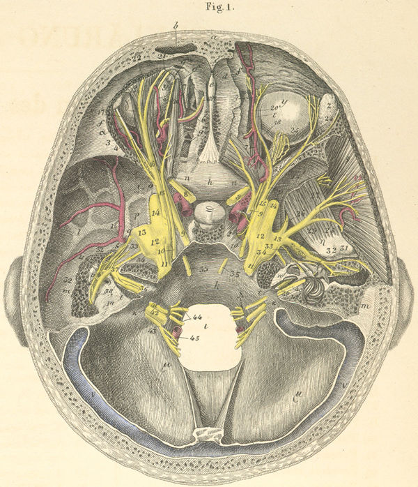
a) Frontal bone.
b) Frontal sinus.
c) Internal frontal spine.
d) Foramen caecum.
e) Crista galli.
f) Frontal bone, orbital portion.
g) Cellulae of the ethmoidal bone.
h) Body of the sphenoid bone.
i) Greater wing of the sphenoid bone.
k) Occipital bone, basilar portion.
l) Temporal bone, squamous portion.
m) Temporal bone, petrous portion.
n) Optic foramen.
o) Foramen rotundum.
p) Foramen ovale.
q) Foramen spinosum.
r) Superior orbital fissure.
s) Tympanic cavity.
t) Internal auditory meatus (broken open from above).
u) Malleus, hammer (s. capitulum).
v) Incus (s. ambos).
w) Cochlea.
x) Superior semicircular canal.
y) Ocular bulb.
z) Lacrimal gland.
a) m. Lateral rectus.
b) m. Levator palpebrae superioris.
g) m. Superior rectus.
d) m. Superior oblique.
d*) Trochlea for superior oblique.
e) m. Medial rectus.
z) m. Temporalis (medial surface).
h) m. Lateral pterygoid.
q) Hypophysis (pituitary gland).
i) Foramen magnum.
k) Jugular foramen.
l) Hypoglossal canal (s. Anterior condyloid
foramen).
m) Occipital bone (fossae cerebelli).
n) Transverse sinus.
Please send us comments by filling out our Comment Form.
All contents copyright © 1995-2025 the Author(s) and Michael P. D'Alessandro, M.D. All rights reserved.
"Anatomy Atlases", the Anatomy Atlases logo, and "A digital library of anatomy information" are all Trademarks of Michael P. D'Alessandro, M.D.
Anatomy Atlases is funded in whole by Michael P. D'Alessandro, M.D. Advertising is not accepted.
Your personal information remains confidential and is not sold, leased, or given to any third party be they reliable or not.
The information contained in Anatomy Atlases is not a substitute for the medical care and advice of your physician. There may be variations in treatment that your physician may recommend based on individual facts and circumstances.
URL: http://www.anatomyatlases.org/