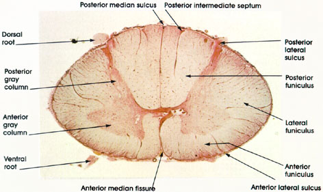

Plate 17.316 Spinal Cord
Ronald A. Bergman, Ph.D., Adel K. Afifi, M.D., Paul M. Heidger,
Jr., Ph.D.
Peer Review Status: Externally Peer Reviewed

Human, Müller's fluid, carmine stain, 8 x.
Dorsal root: The dorsal root carries both myelinated and unmyelinated afferent fibers to the spinal cord. Each fiber is the central process of a dorsal root ganglion cell.
Posterior gray column: Long and narrow column of gray matter reaches almost to the surface of the spinal cord. Primarily concerned with sensory input.
Anterior gray column: Short and broad column of gray matter. Concerned with motor function. Both posterior and anterior gray columns are sites where sensory and motor cell bodies, respectively, are found.
Ventral root: Bundle of somatic motor fibers (axons of somatic motor neurons) and preganglionic fibers (axons of autonomic motor neurons). Constitute the efferent outflow of the spinal cord.
Anterior median fissure: About 3 mm deep. Contains blood vessels (anterior spinal artery) supplying the anterior two thirds of the cord.
Anterior lateral sulcus: Site of exit of ventral root. Hardly distinguishable in this preparation.
Anterior funiculus: Between the anterior median fissure and anterolateral sulcus (ventral root). Merges with the lateral funiculus. Contains ascending and descending tracts.
Lateral funiculus: Between the dorsal and ventral roots. Merges with the anterior funiculus. Contains ascending and descending tracts.
Posterior lateral sulcus: Site of entry of dorsal root.
Posterior funiculus: Between posterior median sulcus and dorsal root. Contains ascending tracts. Posterior intermediate septum: Found only in cervical and upper thoracic segments. Posterior median sullcus: About 5 mm deep, reaches the deep-lying gray matter.
Next Page | Previous Page | Section Top | Title Page
Please send us comments by filling out our Comment Form.
All contents copyright © 1995-2025 the Author(s) and Michael P. D'Alessandro, M.D. All rights reserved.
"Anatomy Atlases", the Anatomy Atlases logo, and "A digital library of anatomy information" are all Trademarks of Michael P. D'Alessandro, M.D.
Anatomy Atlases is funded in whole by Michael P. D'Alessandro, M.D. Advertising is not accepted.
Your personal information remains confidential and is not sold, leased, or given to any third party be they reliable or not.
The information contained in Anatomy Atlases is not a substitute for the medical care and advice of your physician. There may be variations in treatment that your physician may recommend based on individual facts and circumstances.
URL: http://www.anatomyatlases.org/