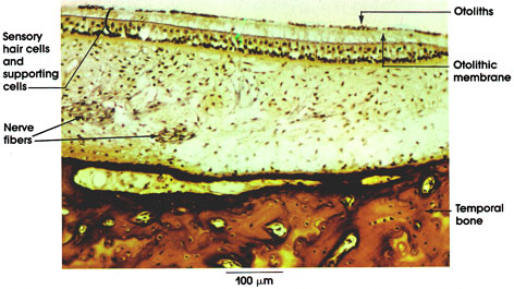

Plate 16.315 Macula Utriculi
Ronald A. Bergman, Ph.D., Adel K. Afifi, M.D., Paul M. Heidger,
Jr., Ph.D.
Peer Review Status: Externally Peer Reviewed

Cat, Müller's fluid, iron hematoxylin, 162 x.
Sensory area of the utricle. The sensory epithelium is composed of two types of cells.
Hair cells: Flask-shaped. Nuclei occupy the upper part of the epithelial sheet. These receptors of the macula utriculi are concerned with the orientation of the body with regard to gravity. The receptor cells of the macula sacculi, however, appear to respond primarily to vibratory stimuli.
Supporting cells: Slender. Nuclei are lined up near the basement membrane of the epithelium.
The surface of the macula is covered by a gelatinous material, the otolithic membrane, through which hairs of the hair cells project. The upper surface of the membrane contains densely packed crystals of a calcium carbonate- protein mixture, the otoliths. The utricle and saccule, which are similar in appearance, constitute the "otolith organ."
Nerve fibers: Afferent components of the vestibular portion of the eighth cranial nerve, the vestibulocochlear nerve.
Temporal bone: Forming the osseous labyrinth.
Next Page | Previous Page | Section Top | Title Page
Please send us comments by filling out our Comment Form.
All contents copyright © 1995-2025 the Author(s) and Michael P. D'Alessandro, M.D. All rights reserved.
"Anatomy Atlases", the Anatomy Atlases logo, and "A digital library of anatomy information" are all Trademarks of Michael P. D'Alessandro, M.D.
Anatomy Atlases is funded in whole by Michael P. D'Alessandro, M.D. Advertising is not accepted.
Your personal information remains confidential and is not sold, leased, or given to any third party be they reliable or not.
The information contained in Anatomy Atlases is not a substitute for the medical care and advice of your physician. There may be variations in treatment that your physician may recommend based on individual facts and circumstances.
URL: http://www.anatomyatlases.org/