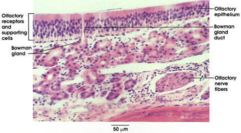

Plate 16.297 Olfactory Mucosa
Ronald A. Bergman, Ph.D., Adel K. Afifi, M.D., Paul M. Heidger,
Jr., Ph.D.
Peer Review Status: Externally Peer Reviewed

Rodent, 10% formalin, H. & E., 218 x.
Olfactory epithelium: Thick pseudostratified epithelium containing (1) sustentacular (supporting) cells, (2) bipolar receptor neurons, and (3) basal cells. Cells are densely packed and difficult to differentiate in thick sections. The nuclei of these cells are layered (from the outside) in the order given above. No goblet cells are located in this region, but the area is flushed by seromucous glands located beneath the epithelium.
Glands of Bowman*: Located in the lamina propria. Branched tubuloalveolar glands that secrete a seromucus. The secretions keep the surface moist, facilitate solution of substances being smelled, and subsequently cleanse the olfactory receptors of the olfactory stimulus.
Olfactory nerve fibers: Non-myelinated axons of bipolar receptor neurons. Nerve bundles are located deep in the lamina propria.
*Bowman was a nineteenth-century English anatomist.
Next Page | Previous Page | Section Top | Title Page
Please send us comments by filling out our Comment Form.
All contents copyright © 1995-2025 the Author(s) and Michael P. D'Alessandro, M.D. All rights reserved.
"Anatomy Atlases", the Anatomy Atlases logo, and "A digital library of anatomy information" are all Trademarks of Michael P. D'Alessandro, M.D.
Anatomy Atlases is funded in whole by Michael P. D'Alessandro, M.D. Advertising is not accepted.
Your personal information remains confidential and is not sold, leased, or given to any third party be they reliable or not.
The information contained in Anatomy Atlases is not a substitute for the medical care and advice of your physician. There may be variations in treatment that your physician may recommend based on individual facts and circumstances.
URL: http://www.anatomyatlases.org/