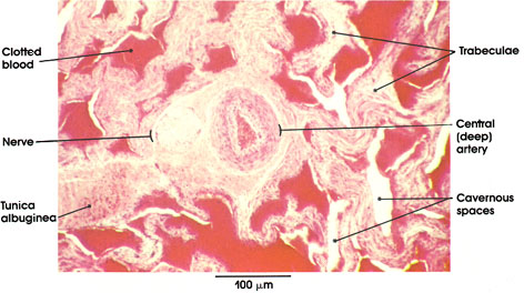

Plate 14.278 Penis
Ronald A. Bergman, Ph.D., Adel K. Afifi, M.D., Paul M. Heidger,
Jr., Ph.D.
Peer Review Status: Externally Peer Reviewed

Human, 10% formalin, H. & E., 162 x.
The erectile tissue of the corpus cavernosum of the penis is composed of cavernous spaces separated by fibromuscular septa or trabeculae. The latter are extensions of the tunica albuginea, the fibrous coat that surrounds the corpus. The cavernous spaces are filled with blood, and the engorgement of these spaces results in the erection of the penis. Note the central (deep) artery, which traverses the corpus cavernosum. This artery gives rise to the spiraling helicine arterioles that open into the sinuses. The central artery is the principal vessel for filling the sinuses during erection.
Adjacent to the central artery, note the nerve cut in cross section. The penis is richly supplied with spinal, sympathetic, and parasympathetic fibers. The autonomic fibers innervate the smooth muscle in the arterial wall and trabeculae.
Next Page | Previous Page | Section Top | Title Page
Please send us comments by filling out our Comment Form.
All contents copyright © 1995-2025 the Author(s) and Michael P. D'Alessandro, M.D. All rights reserved.
"Anatomy Atlases", the Anatomy Atlases logo, and "A digital library of anatomy information" are all Trademarks of Michael P. D'Alessandro, M.D.
Anatomy Atlases is funded in whole by Michael P. D'Alessandro, M.D. Advertising is not accepted.
Your personal information remains confidential and is not sold, leased, or given to any third party be they reliable or not.
The information contained in Anatomy Atlases is not a substitute for the medical care and advice of your physician. There may be variations in treatment that your physician may recommend based on individual facts and circumstances.
URL: http://www.anatomyatlases.org/