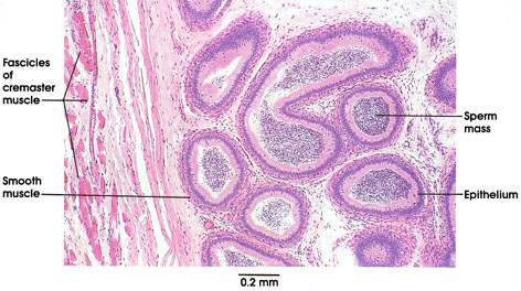

Plate 14.270 Epididymis
Ronald A. Bergman, Ph.D., Adel K. Afifi, M.D., Paul M. Heidger,
Jr., Ph.D.
Peer Review Status: Externally Peer Reviewed

Human, 10% formalin, H. & E., 54 x.
The epididymis, a highly coiled duct approximately 6 m in length in man, is firmly adherent to the gonad. in histological preparations, therefore, it is not unusual to observe sections of both testis and epididymis. The duct is divided anatomically into three major portions (head, body, and tail) and by histological criteria, several further subdivisions may be recognized. Because of the tight coiling of the duct, histological sections invariably reveal many tubular profiles in different planes of section.
The portion of the epididymis shown here is from the distal portion of the head, in which the epithelial lining of the duct is a tall pseudostratified columnar epithelium, lacking goblet cells. The tall columnar cells bear prominent (non-motile) stereocilia on their apical surfaces, facilitating the resorption of testicular tubular fluid in which the spermatozoa are transported to the organ. A thin coat of smooth muscle surrounds the duct and is readily differentiated from the loose connective tissue surrounding the coils of the duct. A packed mass of non-motile sperm is seen within the lumen of the epididymis. In this preparation, striated muscle fibers belonging to the cremaster muscle are seen investing the organ. This muscle of the spermatic cord enables the gonad to be retracted within the scrotum toward the, abdomen. A higher power view of the epididymis is seen in Plate 271.
Next Page | Previous Page | Section Top | Title Page
Please send us comments by filling out our Comment Form.
All contents copyright © 1995-2025 the Author(s) and Michael P. D'Alessandro, M.D. All rights reserved.
"Anatomy Atlases", the Anatomy Atlases logo, and "A digital library of anatomy information" are all Trademarks of Michael P. D'Alessandro, M.D.
Anatomy Atlases is funded in whole by Michael P. D'Alessandro, M.D. Advertising is not accepted.
Your personal information remains confidential and is not sold, leased, or given to any third party be they reliable or not.
The information contained in Anatomy Atlases is not a substitute for the medical care and advice of your physician. There may be variations in treatment that your physician may recommend based on individual facts and circumstances.
URL: http://www.anatomyatlases.org/