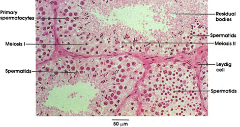

Plate 14.267 Testis
Ronald A. Bergman, Ph.D., Adel K. Afifi, M.D., Paul M. Heidger,
Jr., Ph.D.
Peer Review Status: Externally Peer Reviewed

Monkey, glutaraldehyde, 1.5 µm plastic section, H. & E., 216 x.
The process of spermatogenesis is best appreciated from the study of the various stages represented in adjacent sections of a seminiferous tubule. Occasionally, however, a tubule is cut in fortuitous tangential section in such a manner that the processes occurring along its length can be appreciated. Such is the case in this figure, in which, toward the upper left of the section, primary spermatocytes are seen; these are the daughter cells of the basally situated spermatogonia. Note their distinctive meiotic prophase nuclei. Division of primary spermatocytes (meiosis 1) gives rise to daughter cells, the secondary spermatocytes. These, in turn, complete the second meiotic division, giving rise to haploid daughter cells, the round spermatids (upper right). Over time, the round spermatids complete cytodifferentiation and become mature, elongate spermatids. These cells lie most apically within the epithelium, among the cytoplasmic residual bodies. Mature spermatids shed into the tubular lumen are termed spermatozoa. Other tubules (such as the one in the lower right portion of the figure) may be cross-sectioned tangentially such that the plane of section passes predominantly through one cell population of the seminiferous epithelium. This accounts for the absence of a luminal profile in this tubule and the predominance of round spermatids.
Next Page | Previous Page | Section Top | Title Page
Please send us comments by filling out our Comment Form.
All contents copyright © 1995-2025 the Author(s) and Michael P. D'Alessandro, M.D. All rights reserved.
"Anatomy Atlases", the Anatomy Atlases logo, and "A digital library of anatomy information" are all Trademarks of Michael P. D'Alessandro, M.D.
Anatomy Atlases is funded in whole by Michael P. D'Alessandro, M.D. Advertising is not accepted.
Your personal information remains confidential and is not sold, leased, or given to any third party be they reliable or not.
The information contained in Anatomy Atlases is not a substitute for the medical care and advice of your physician. There may be variations in treatment that your physician may recommend based on individual facts and circumstances.
URL: http://www.anatomyatlases.org/