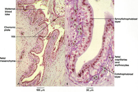

Plate 13.259 Placenta
Ronald A. Bergman, Ph.D., Adel K. Afifi, M.D., Paul M. Heidger,
Jr., Ph.D.
Peer Review Status: Externally Peer Reviewed

Human, Bouin's-Halmi's AFT, A. 121 x; B. 357 x.
Fetal components of the placenta include the chorionic plate and the villi that originate from it (A); a high-magnification view of the epithelial lining of these structures is shown in B. In A, note that the epithelium-lined chorionic plate gives rise to villi, which project into the maternal blood space. Fetal mesenchyme constitutes the core of the villi; fetal blood vessels are present in the plate, and a rich capillary network is formed within the villi, analogous to the histophysiological arrangement of vessels within intestinal villi, which are also specialized to optimize absorptive capacity. Note that the capillaries in B contain fetal erythrocytes (stained green), which contain nuclei. The villi are lined by two layers of embryonic trophoblast. The inner cytotrophoblast consists of cells that give rise to the outer syncytial layer, the syncytiotroph o blast. Gradually, during the latter half of pregnancy, the cytotrophoblast is largely incorporated within the syncytial layer; therefore, specimens obtained at delivery will demonstrate only the syncytial layer. This histological feature can assume importance to the forensic pathologist in medicolegal examination of placental specimens, as well as to inexperienced histologists seeking a layer that no longer exists in the most common source of placental tissues, the afterbirth.
Next Page | Previous Page | Section Top | Title Page
Please send us comments by filling out our Comment Form.
All contents copyright © 1995-2025 the Author(s) and Michael P. D'Alessandro, M.D. All rights reserved.
"Anatomy Atlases", the Anatomy Atlases logo, and "A digital library of anatomy information" are all Trademarks of Michael P. D'Alessandro, M.D.
Anatomy Atlases is funded in whole by Michael P. D'Alessandro, M.D. Advertising is not accepted.
Your personal information remains confidential and is not sold, leased, or given to any third party be they reliable or not.
The information contained in Anatomy Atlases is not a substitute for the medical care and advice of your physician. There may be variations in treatment that your physician may recommend based on individual facts and circumstances.
URL: http://www.anatomyatlases.org/