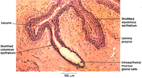

Plate 12.244 Urethra
Ronald A. Bergman, Ph.D., Adel K. Afifi, M.D., Paul M. Heidger,
Jr., Ph.D.
Peer Review Status: Externally Peer Reviewed

Human, 10% formalin, H. & E., 162 x.
This plate shows the histology of the Cavernous portion of the male urethra. This portion of the urethra extends throughout the penis to open at the end of the glans. Note the stratified columnar epithelium mucosal lining intermixed with stratified squamous epithelium. The latter type of epithelium is found in interrupted areas throughout the extent of the urethra and is the only epithelial type found at the external opening of the urethra.
Note the deep recesses of the mucosal surface known as lacunae of Morgagni* Isolated intraepithelial mucous gland cells are seen interspersed between the stratified columnar cells lining the lacunae.
The lamina propria is made up of loose connective tissue rich in elastic fibers.
*Morgagni was an eighteenth-century Italian anatomist and pathologist.
Next Page | Previous Page | Section Top | Title Page
Please send us comments by filling out our Comment Form.
All contents copyright © 1995-2025 the Author(s) and Michael P. D'Alessandro, M.D. All rights reserved.
"Anatomy Atlases", the Anatomy Atlases logo, and "A digital library of anatomy information" are all Trademarks of Michael P. D'Alessandro, M.D.
Anatomy Atlases is funded in whole by Michael P. D'Alessandro, M.D. Advertising is not accepted.
Your personal information remains confidential and is not sold, leased, or given to any third party be they reliable or not.
The information contained in Anatomy Atlases is not a substitute for the medical care and advice of your physician. There may be variations in treatment that your physician may recommend based on individual facts and circumstances.
URL: http://www.anatomyatlases.org/