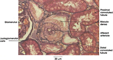

Plate 12.237 Kidney: Cortex
Ronald A. Bergman, Ph.D., Adel K. Afifi, M.D., Paul M. Heidger,
Jr., Ph.D.
Peer Review Status: Externally Peer Reviewed

Rhesus monkey, Zenker's fluid, Mallory's stain, 612 x.
Afferent arteriole: Seen here in cross section. Its proximity to the glomerulus (not seen here), which it serves, is indicated by the presence of cells containing conspicuous granules, which replace smooth muscle fibers normally found in the wall of arterioles.
Juxtaglomerular cells: Rich in cytoplasmic granules containing renin. Renin secreted into the blood is known to play a role in the formation of a hypertensive substance known as angiotensin II.
Glomerulus: Tuft of capillaries having their origin from the afferent arteriole and surrounded by Bowman's capsule. The glomerulus, together with Bowman's capsule, constitutes the renal corpuscle.
Proximal convoluted tubule: Outlet of Bowman's capsule approximately 14 mm long with many small loops near the renal corpuscle. It ultimately straightens and runs toward the medulla in the medullary rays.
Distal convoluted tubule: This portion of the renal tubule has many short loops in close association with the proximal convoluted tubule and the glomerulus. It is about one third the length of the proximal tubule. The distal tubule is continuous with the collecting tubules.
Macula densa: Specialized region of the distal convoluted tubule with tightly packed tubule cells in contact with the afferent arteriole. The bases of the cells of the macula densa are consistently found in intimate association with the juxtaglomerular cells in the wall of the afferent arteriole. Because this structural relationship suggests a functional relationship (supported experimentally), the macula densa and the juxtaglomerular cells together are referred to as the juxtaglomerular apparatus.
Next Page | Previous Page | Section Top | Title Page
Please send us comments by filling out our Comment Form.
All contents copyright © 1995-2025 the Author(s) and Michael P. D'Alessandro, M.D. All rights reserved.
"Anatomy Atlases", the Anatomy Atlases logo, and "A digital library of anatomy information" are all Trademarks of Michael P. D'Alessandro, M.D.
Anatomy Atlases is funded in whole by Michael P. D'Alessandro, M.D. Advertising is not accepted.
Your personal information remains confidential and is not sold, leased, or given to any third party be they reliable or not.
The information contained in Anatomy Atlases is not a substitute for the medical care and advice of your physician. There may be variations in treatment that your physician may recommend based on individual facts and circumstances.
URL: http://www.anatomyatlases.org/