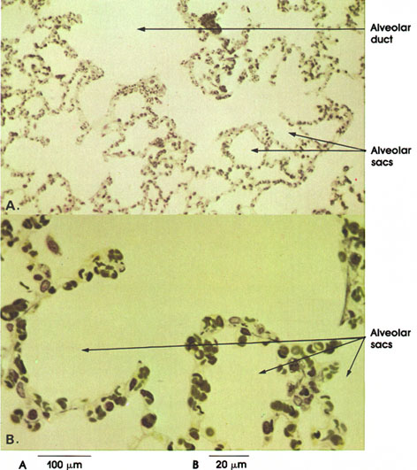

Plate 11.229 Alviolar Duct and Alveolar Sacs
Ronald A. Bergman, Ph.D., Adel K. Afifi, M.D., Paul M. Heidger,
Jr., Ph.D.
Peer Review Status: Externally Peer Reviewed

Rat, glutaraldehyde-osmium fixation,
toluidine blue stain, A. 162 x, B. 612 x.
Refer to Figure 11A which can be used in conjunction with this plate in order to follow the structural changes that occur from the respiratory bronchiole to alveolar ducts to alveolar sacs where gaseous exchange takes place.
Alveolar duct: A branch of the respiratory bronchiole. The duct is composed of alveolar sacs and alveoli. See Plates 227 and 228.
Alveolar sacs: Cluster of alveoli opening into the main lumen of the sac. Individual alveoli are lined by a thin squamous (Pneumocyte type I) and cuboidal epithelium (Pneumocyte type II). A closely applied capillary network is separated from the epithelium by a thin basement membrane. It is across this trilaminar wall that oxygen, carbon dioxide and other inspired gases are exchanged in respiration.
Next Page | Previous Page | Section Top | Title Page
Please send us comments by filling out our Comment Form.
All contents copyright © 1995-2025 the Author(s) and Michael P. D'Alessandro, M.D. All rights reserved.
"Anatomy Atlases", the Anatomy Atlases logo, and "A digital library of anatomy information" are all Trademarks of Michael P. D'Alessandro, M.D.
Anatomy Atlases is funded in whole by Michael P. D'Alessandro, M.D. Advertising is not accepted.
Your personal information remains confidential and is not sold, leased, or given to any third party be they reliable or not.
The information contained in Anatomy Atlases is not a substitute for the medical care and advice of your physician. There may be variations in treatment that your physician may recommend based on individual facts and circumstances.
URL: http://www.anatomyatlases.org/