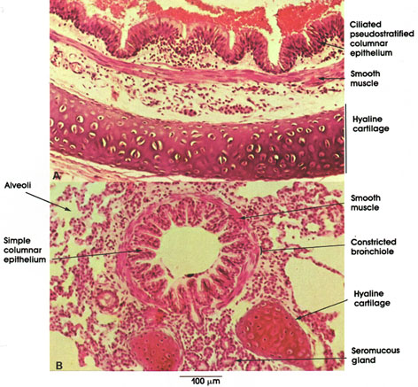

Plate 11.225 Bronchus and Bronchiole
Ronald A. Bergman, Ph.D., Adel K. Afifi, M.D., Paul M. Heidger,
Jr., Ph.D.
Peer Review Status: Externally Peer Reviewed

Cat, 10% formalin, H. & E. stain, 162 x.
In A, part of a bronchus can be seen. Bronchi are lined by pseudostratified columnar epithelium with goblet cells. The thickness and layering of the epithelium decreases gradually with the decrease in size of bronchi. A smooth muscle layer encircles a thin connective tissue lamina propria. In contrast to the trachea, the smooth muscle of the bronchus is arranged in interlacing spirals around the bronchus. Between the smooth muscle layer and the cartilage is the submucosa, which may contain seromucous glands, not seen in this preparation. The hyaline cartilage is arranged in discontinuous plates around the bronchus.
In B, a bronchiole is seen in the midst of respiratory tissue. The epithelium is simple columnar ciliated. The lamina propria is replaced by the muscle layer that encircles the bronchiole. in man, cartilage and glands are characteristically present until bronchioles decrease in size to approximately 0.5 mm in diameter.
Next Page | Previous Page | Section Top | Title Page
Please send us comments by filling out our Comment Form.
All contents copyright © 1995-2025 the Author(s) and Michael P. D'Alessandro, M.D. All rights reserved.
"Anatomy Atlases", the Anatomy Atlases logo, and "A digital library of anatomy information" are all Trademarks of Michael P. D'Alessandro, M.D.
Anatomy Atlases is funded in whole by Michael P. D'Alessandro, M.D. Advertising is not accepted.
Your personal information remains confidential and is not sold, leased, or given to any third party be they reliable or not.
The information contained in Anatomy Atlases is not a substitute for the medical care and advice of your physician. There may be variations in treatment that your physician may recommend based on individual facts and circumstances.
URL: http://www.anatomyatlases.org/