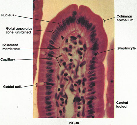

Plate 10.194 Duodenum
Ronald A. Bergman, Ph.D., Adel K. Afifi, M.D., Paul M. Heidger,
Jr., Ph.D.
Peer Review Status: Externally Peer Reviewed

Cat, Helly's fluid, H. & E., 612 x.
The word villus is of Latin origin, meaning shaggy hair or a tuft of hair. The intestinal villi project from the intestinal wall like hairs or the nap on cloth. The term villus was first coined for the intestinal projections by Berengarius, an Italian anatomist, in 1524.
Columnar epithelium: Covers the surface of the villus. Surface of the epithelium has a striated border (microvilli by electron microscopy) to increase its absorptive surface. Products obtained from the extracellular digestive process, salts, vitamins, and other substances are carried through the cytoplasm of these cells and delivered to the connective tissue to enter the blood vessels or lymphatics. The surface epithelia[ cells are being continuously shed from the apex of the villus (extrusion zone) and replaced by migrating cells from the bottom of the crypts (Plates 29 and 198).
Nucleus: Ovoid, located in the lower half of the columnar cell.
Golgi apparatus zone: Relatively pale area in this preparation. Specific stains are needed to demonstrate the Golgi apparatus, which lies between the nuclei and free surface.
Lymphocytes: One of the cell types commonly found in the lamina propria. Seen migrating into the epithelial layer to be extruded into the lumen. See Plates 29 and 198.
Goblet cell: Dispersed among the columnar absorptive epithelial cells. They appear empty because some mucin is lost during the preparation of the specimen. The residual mucin stains poorly with the H. & E. stain. Nucleus is basally located. Compare the small number of goblet cells in this preparation with their abundant number in another region of the intestine (Plate 207).
Basement membrane: A delicate membrane that supports the epithelium. Composed primarily of reticular fibers embedded in an amorphous protein polysaccharide ground substance.
Central lacteal: A lymph vessel situated near the center of the villus. Note its endothelial lining. The lacteals become distended during absorption of fat.
Capillary: Capillaries of the villus form a network that lies underneath the basement membrane of the lining epithelium.
Next Page | Previous Page | Section Top | Title Page
Please send us comments by filling out our Comment Form.
All contents copyright © 1995-2025 the Author(s) and Michael P. D'Alessandro, M.D. All rights reserved.
"Anatomy Atlases", the Anatomy Atlases logo, and "A digital library of anatomy information" are all Trademarks of Michael P. D'Alessandro, M.D.
Anatomy Atlases is funded in whole by Michael P. D'Alessandro, M.D. Advertising is not accepted.
Your personal information remains confidential and is not sold, leased, or given to any third party be they reliable or not.
The information contained in Anatomy Atlases is not a substitute for the medical care and advice of your physician. There may be variations in treatment that your physician may recommend based on individual facts and circumstances.
URL: http://www.anatomyatlases.org/