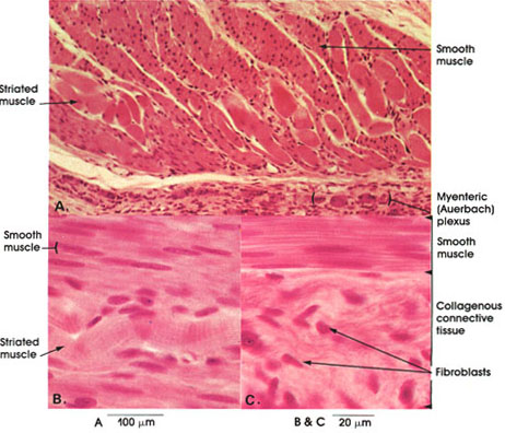

Plate 10.188 Esophagus
Ronald A. Bergman, Ph.D., Adel K. Afifi, M.D., Paul M. Heidger,
Jr., Ph.D.
Peer Review Status: Externally Peer Reviewed

Dog, 10% formalin, H. & E., A. 162 x., B. & C. 612 x.
The external muscular coat of the esophagus is made up entirely of skeletal muscles in the upper third, of skeletal and smooth muscles in the middle third (A and B), and of purely smooth muscle in the lower third (C). Outside the muscle layer is a layer of collagenous connective tissue with fibroblasts, the adventitia (C) The orientation of muscle fibers also varies. Typically, an inner circular and outer longitudinal layer exist, but many bundles are arranged obliquely or in a spiral fashion. Between the two layers of muscle is a nerve plexus associated with numerous small ganglia, the myenteric plexus of Auerbach (A). This is mainly a parasympathetic (vagus nerve) plexus along with some postganglionic sympathetic nerves. it is named after Leopold Auerbach, a German anatomist, who described it in 1862.
Next Page | Previous Page | Section Top | Title Page
Please send us comments by filling out our Comment Form.
All contents copyright © 1995-2025 the Author(s) and Michael P. D'Alessandro, M.D. All rights reserved.
"Anatomy Atlases", the Anatomy Atlases logo, and "A digital library of anatomy information" are all Trademarks of Michael P. D'Alessandro, M.D.
Anatomy Atlases is funded in whole by Michael P. D'Alessandro, M.D. Advertising is not accepted.
Your personal information remains confidential and is not sold, leased, or given to any third party be they reliable or not.
The information contained in Anatomy Atlases is not a substitute for the medical care and advice of your physician. There may be variations in treatment that your physician may recommend based on individual facts and circumstances.
URL: http://www.anatomyatlases.org/