
Ronald A. Bergman, Ph.D., Adel K. Afifi, M.D., Paul M. Heidger,
Jr., Ph.D.
Peer Review Status: Externally Peer Reviewed

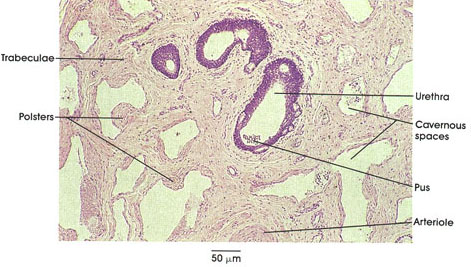
Human, 10% formalin, H. & E., 187 x.
Cavernous tissues (the spongiosum and paired cavernous bodies of clitoris and penis) increase in size by filling with blood and change a flaccid organ to a rigid one. Cavernous organs are filled with a complex network of venous sinuses separated by trabeculae composed of smooth muscle and connective tissue.
The spaces and trabeculae are lined with endothleium. Note that the walls of cavernous tissues possess subendothelial thickenings composed of longitudinal muscle fibers called polsters, which partially occlude the lumen of the sinus. it is believed that the polsters facilitate the closure of sinuses during erection. Note also the pseudostratified columnar epithelium of the urethral diverticula with their unique intraepithelial mucous gland cells (glands of Littré*), which can be seen in this illustration.
*Littré was an eighteenth-century French (Paris) anatomist.
Next Page | Previous Page | Section Top | Title Page
Please send us comments by filling out our Comment Form.
All contents copyright © 1995-2025 the Author(s) and Michael P. D'Alessandro, M.D. All rights reserved.
"Anatomy Atlases", the Anatomy Atlases logo, and "A digital library of anatomy information" are all Trademarks of Michael P. D'Alessandro, M.D.
Anatomy Atlases is funded in whole by Michael P. D'Alessandro, M.D. Advertising is not accepted.
Your personal information remains confidential and is not sold, leased, or given to any third party be they reliable or not.
The information contained in Anatomy Atlases is not a substitute for the medical care and advice of your physician. There may be variations in treatment that your physician may recommend based on individual facts and circumstances.
URL: http://www.anatomyatlases.org/