
Ronald A. Bergman, Ph.D., Adel K. Afifi, M.D., Paul M. Heidger,
Jr., Ph.D.
Peer Review Status: Externally Peer Reviewed

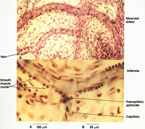
Rabbit, 10% formalin, hematoxylin stain, A. 162 x., B. 612 x.
This plate is taken of a thin spread of mesentery showing the configuration of blood vessels. Compare the size of the muscular artery and vein in A. Note the thicker wall of the artery and the circular bands of smooth muscles in the wall. In B, a row of smooth muscle nuclei is seen, circumferentially arranged in the wall of the arteriole. Their elongated form is not seen because they are shown here in only two dimensions. Note the precapillary sphincter at the junction of an arteriole and capillary. The precapillaries are larger than ordinary capillaries and contain smooth muscle fibers that encircle the vessel and act as sphincters to control blood flow in the capillary bed. Note the absence of smooth muscle nuclei from the capillary. The nuclei oriented along the long axis of the capillary belong to the endothelial cells.
Next Page | Previous Page | Section Top | Title Page
Please send us comments by filling out our Comment Form.
All contents copyright © 1995-2025 the Author(s) and Michael P. D'Alessandro, M.D. All rights reserved.
"Anatomy Atlases", the Anatomy Atlases logo, and "A digital library of anatomy information" are all Trademarks of Michael P. D'Alessandro, M.D.
Anatomy Atlases is funded in whole by Michael P. D'Alessandro, M.D. Advertising is not accepted.
Your personal information remains confidential and is not sold, leased, or given to any third party be they reliable or not.
The information contained in Anatomy Atlases is not a substitute for the medical care and advice of your physician. There may be variations in treatment that your physician may recommend based on individual facts and circumstances.
URL: http://www.anatomyatlases.org/