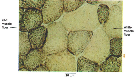

Semitendinosus, cross section
Mitochondria; succinic dehydrogenase localization
Ronald A. Bergman, Ph.D., Adel K. Afifi, M.D., Paul M. Heidger,
Jr., Ph.D.
Peer Review Status: Externally Peer Reviewed

Rat, frozen section, Tetrazolium method, 612 x.
Histochemical methods similar to the one used here, which is specific for mitochrondria, have been instrumental in distinguishing muscle fiber types in health and disease.
Red muscle fiber (B fiber): Rich in mitochondria and lipids, this type of fiber is slow contracting. Known in the human as Type I muscle fiber.
White muscle fiber (A fiber): Relatively poor in mitochondria and lipids, but rich in myofibrillar ATPase activity and glycogen, these fibers are fast contracting. Known in the human as Type II muscle fiber.
Next Page | Previous Page | Section Top | Title Page
Please send us comments by filling out our Comment Form.
All contents copyright © 1995-2025 the Author(s) and Michael P. D'Alessandro, M.D. All rights reserved.
"Anatomy Atlases", the Anatomy Atlases logo, and "A digital library of anatomy information" are all Trademarks of Michael P. D'Alessandro, M.D.
Anatomy Atlases is funded in whole by Michael P. D'Alessandro, M.D. Advertising is not accepted.
Your personal information remains confidential and is not sold, leased, or given to any third party be they reliable or not.
The information contained in Anatomy Atlases is not a substitute for the medical care and advice of your physician. There may be variations in treatment that your physician may recommend based on individual facts and circumstances.
URL: http://www.anatomyatlases.org/