
Developing eosinophils
Ronald A. Bergman, Ph.D., Adel K. Afifi, M.D., Paul M. Heidger,
Jr., Ph.D.
Peer Review Status: Externally Peer Reviewed

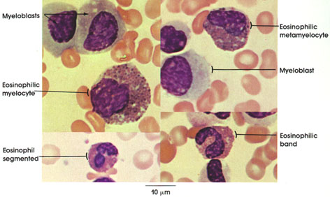
Human, air-dried marrow smear, Wright's stain, 1416 x.
Myeloblast: Stem cell of the leucocytic series. It has a rounded large nucleus and lightly basophilic agranular cytoplasm. See also Plate 58.
Eosinophilic myelocyte: These develop from myeloblasts. Specific acidophilic granules appear in cytoplasm. The nucleus is rounded or oval. Chromatin of nucleus is coarser than in the myeloblast. This cell is capable of division.
Eosinophilic metamyelocyte: This cell is no longer capable of cell division. The nucleus is kidney- shaped or indented. Cytoplasm contains acidophilic granules.
Eosinophilic band: Immature or juvenile eosinophil. The nucleus is horseshoe- or drumstick-shaped, and there are eosinophilic granules in cytoplasm.
Segmented eosinophil: Mature eosinophil. The nucleus is lobulated and the lobes are connected with thin chromatin threads. There is abundant granular cytoplasm.
Next Page | Previous Page | Section Top | Title Page
Please send us comments by filling out our Comment Form.
All contents copyright © 1995-2025 the Author(s) and Michael P. D'Alessandro, M.D. All rights reserved.
"Anatomy Atlases", the Anatomy Atlases logo, and "A digital library of anatomy information" are all Trademarks of Michael P. D'Alessandro, M.D.
Anatomy Atlases is funded in whole by Michael P. D'Alessandro, M.D. Advertising is not accepted.
Your personal information remains confidential and is not sold, leased, or given to any third party be they reliable or not.
The information contained in Anatomy Atlases is not a substitute for the medical care and advice of your physician. There may be variations in treatment that your physician may recommend based on individual facts and circumstances.
URL: http://www.anatomyatlases.org/