
Stomach
fundus
Ronald A. Bergman, Ph.D., Adel K. Afifi, M.D., Paul M. Heidger,
Jr., Ph.D.
Peer Review Status: Externally Peer Reviewed

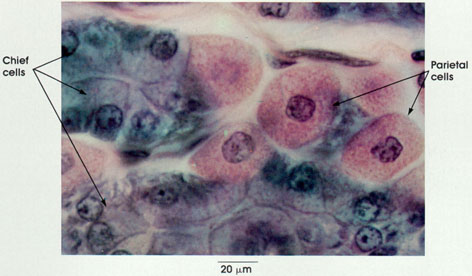
Dog, Helly's fluid, H. & E., 612 x.
Cells whose cytoplasm stains intensely blue with hematoxylin are primarily involved in protein synthesis (e.g., chief cells). Cytoplasm that is eosinophilic (stained with eosin) contains little RNA and is engaged in other kinds of cellular activity (e.g., muscular contraction, active transport, or the elaboration of hydrochloric acid, as in this preparation). See Appendix IV for a brief discussion of the staining mechanism and the hematoxylin and eosin dyes.
Chief cells: Pyramidal shape. Basal nucleus. Granular apex contains zymogen granules. Basophilia of base is due to cytoplasmic ribonucleic acid (RNA). Secretes pepsinogen and rennin.
Parietal cells: Wedged between and larger than chief cells. Centrally placed nuclei surrounded by a finely granular acidophilic cytoplasm. Cytoplasm granularity is due to abundant but unstained mitochondria. Secrete hydrochloric acid, which converts pepsinogen to pepsin, a proteolytic digestive enzyme active at very low pH and is the source of the antipernicious anemia factor in humans. See Plates 190 and 191.
Next Page | Previous Page | Section Top | Title Page
Please send us comments by filling out our Comment Form.
All contents copyright © 1995-2025 the Author(s) and Michael P. D'Alessandro, M.D. All rights reserved.
"Anatomy Atlases", the Anatomy Atlases logo, and "A digital library of anatomy information" are all Trademarks of Michael P. D'Alessandro, M.D.
Anatomy Atlases is funded in whole by Michael P. D'Alessandro, M.D. Advertising is not accepted.
Your personal information remains confidential and is not sold, leased, or given to any third party be they reliable or not.
The information contained in Anatomy Atlases is not a substitute for the medical care and advice of your physician. There may be variations in treatment that your physician may recommend based on individual facts and circumstances.
URL: http://www.anatomyatlases.org/