
Illustrated Encyclopedia of Human Anatomic Variation: Opus III: Nervous System
Ronald A. Bergman, PhD
Adel K. Afifi, MD, MS
Ryosuke Miyauchi, MD
Peer Review Status: Internally Peer Reviewed
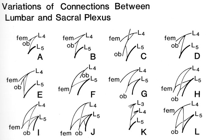 The connections between the lumbar and the sacral plexus
were examined bilaterally in 122 subjects. In 91.8% of subjects the
plexuses were joined, by a single nervus furcalis and in 0.8% by a
doubled nervus furcalis. The single furcal nerve was formed by the
abdominal nerve L4 in 80% and L5 in 7.7% of cases. The doubled furcal
nerve arose from L3 and L4 in 0.4% of cases and from L4 and L5 in
0.4%. Usually, the major part of the furcal nerve arising from L4
went to the lumbar plexus, and from L5 to the sacral plexus. In 7.4%
of cases no connection between the plexuses could be found.
The connections between the lumbar and the sacral plexus
were examined bilaterally in 122 subjects. In 91.8% of subjects the
plexuses were joined, by a single nervus furcalis and in 0.8% by a
doubled nervus furcalis. The single furcal nerve was formed by the
abdominal nerve L4 in 80% and L5 in 7.7% of cases. The doubled furcal
nerve arose from L3 and L4 in 0.4% of cases and from L4 and L5 in
0.4%. Usually, the major part of the furcal nerve arising from L4
went to the lumbar plexus, and from L5 to the sacral plexus. In 7.4%
of cases no connection between the plexuses could be found.
Authors' note: Urbanowiez considers illustrations A through E be type I variations, F through J to be type II, K to be type III, and L to be type IV
L3 to L5, respective lumbar nerves; fem, femoral nerve; ob, obturator nerve.
Redrawn from Urbanowicz, Z. Connections between the lumbar and the sacral plexus in man. Folis Morphol. 40:271-279, 1981.
Section Top | Title Page
Please send us comments by filling out our Comment Form.
Anatomy Atlases is licensed under a Creative Commons Attribution-NonCommercial-ShareAlike 4.0 International License. 
"Anatomy Atlases", the Anatomy Atlases logo, and "A digital library of anatomy information" are all Trademarks of Michael P. D'Alessandro, M.D.
Anatomy Atlases is funded in whole by Michael P. D'Alessandro, M.D. Advertising is not accepted.
Your personal information remains confidential and is not sold, leased, or given to any third party be they reliable or not.
The information contained in Anatomy Atlases is not a substitute for the medical care and advice of your physician. There may be variations in treatment that your physician may recommend based on individual facts and circumstances.
URL: http://www.anatomyatlases.org/