
Illustrated Encyclopedia of Human Anatomic Variation: Opus II: Cardiovascular System
Ronald A. Bergman, PhD
Adel K. Afifi, MD, MS
Ryosuke Miyauchi, MD
Peer Review Status: Internally Peer Reviewed

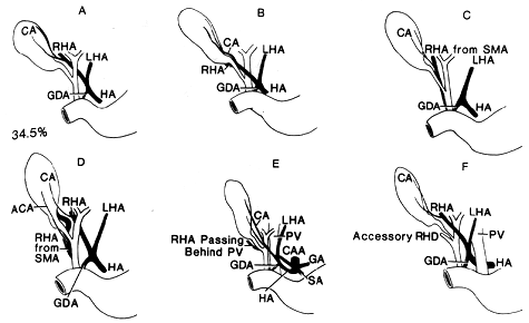
The arrangement of the vessels and ducts given as normal constitute only 69 cases of the 200 studied (34.5%) by Flint (A). "So frequent are variations that it is impossible to regard any one type as normal; the arrangement found in the 69 cases can only be described as the most usual one."
ACA, Accessory cystic artery; CA, cystic artery; CAA, celiac trunk; CD, cystic duct; GA, gastric artery; GDA, gastroduodenal artery; HA, hepatic artery; LHA, left hepatic artery; PV, portal vein; RHA, right hepatic artery; RHD, right hepatic duct; SA, splenic artery; SMA, superior mesenteric artery; SPDA, superior pancreatoduodenal artery.
Right Hepatic Artery
This artery arose from the common hepatic in 158 cases, and reached
the liver by passing behind the common hepatic duct in 136 cases (A),
and ih front of the duct in 22 cases (B). In 42 cases the right
hepatic artery arose from the superior mesenteric (C), and always
passed behind the common duct. In 7 cases there were two right
hepatic arteries, one from the hepatic trunk and one from the
superior mesenteric artery (D). In 2 cases there were two right
hepatic arteries both from the common hepatic, one passed in front
of, and the other behind, the common hepatic duct.
In 4 cases, in addition to passing behind the ducts, the common hepatic or the right hepatic artery also passed behind the portal vein (E and W).
Authors' note: The right hepatic artery may also arise from the aorta, right renal, gastric, or inferior mesenteric artery. No such cases were found in Flint's series.
Cystic Artery
The cystic artery arose from the right hepatic artery in 196 of the
200 cases; in 3 cases from the left hepatic (I and Y), and in 1 case
from the gastroduodenal artery (H).
In 32 cases it passed in front of ihe common hepatic duct (E and G), and in 168 it arose from the right side of the common hepatic duct (A) or behind it (A'), the former being the more common.
Accessory Cystic Artery
In 31 cases there was an accessory cystic artery and in 169 cases a
single cystic. The accessory cystic artery arose from the right
hepatic in 16 (D, J, and K), from the left hepatic in 3 (L and Y),
from the gastroduodional in 11 (M), and from the superior
pancreatoduodenal in 1 case (N) out of the 200.
Left Hepatic Artery (100 cases studied)
In 32 cases there were two left hepatic arteries with one coming from
the common hepatic and the other from the gastric, and in 1 case the
left lobe of the liver received its arterial supply from the gastric
only.
Bile Ducts
According to Flint, the most common point at which union actually
occurs is within 1 cm of the upper border of the duodenum (H). In 28
cases there were no supraduodenal common bile ducts at all (T), the
union taking place at a point anywhere from behind the upper border
of the duodenum to the part embedded in the pancreas, and in 3 cases
the only representative of the common duct was the part that lies in
the wall of the duodenum (U).
Accessory Bile Ducts
Flint reported 29 examples of accessory right hepatic ducts (F, X, Y,
A', and C').
Redrawn from Flint, E.F. Abnormalities of the right hepatic, cystic, and gastroduodenal arteries, and of the bile ducts. Br. J. Surg. 10:509-519, 1923-24.
Section Top | Title Page
Please send us comments by filling out our Comment Form.
All contents copyright © 1995-2025 the Author(s) and Michael P. D'Alessandro, M.D. All rights reserved.
"Anatomy Atlases", the Anatomy Atlases logo, and "A digital library of anatomy information" are all Trademarks of Michael P. D'Alessandro, M.D.
Anatomy Atlases is funded in whole by Michael P. D'Alessandro, M.D. Advertising is not accepted.
Your personal information remains confidential and is not sold, leased, or given to any third party be they reliable or not.
The information contained in Anatomy Atlases is not a substitute for the medical care and advice of your physician. There may be variations in treatment that your physician may recommend based on individual facts and circumstances.
URL: http://www.anatomyatlases.org/